Home
/ Medial Femoral Condyle, Eorthopod Patient Education : If there is a fracture (break) in part of the condyle, this is known as a fracture of the femoral condyle.
Medial Femoral Condyle, Eorthopod Patient Education : If there is a fracture (break) in part of the condyle, this is known as a fracture of the femoral condyle.
Medial Femoral Condyle, Eorthopod Patient Education : If there is a fracture (break) in part of the condyle, this is known as a fracture of the femoral condyle.. Juvenile ocd lesions have a better healing prognosis than adults. Sagittal plane fracture of the medial femoral condyle. This adaptable graft option is most commonly used with oats or shell techniques to restore cartilage in the knee, but can also be used to for cartilage restoration of other joints. This is associated with a positive bone scan and, frequently, a radiolucent lesion in the subchondral zone. Of the six patients who had suffered an isolated fracture of their medial condyle, four of the patients had their fractures diagnosed on the first visit.
This adaptable graft option is most commonly used with oats or shell techniques to restore cartilage in the knee, but can also be used to for cartilage restoration of other joints. Your knee mri will often show a: The femoral condyle is a thickened area of the femur just above the knee. The inner side of the knee or the medial femoral condyle is the most common area for a cartilage defect. An imbalance of the muscles around the knee (some muscles are weaker than others.) overuse (repeated bending or twisting) of the knee joint, especially during sports.
Medial Femoral Condyle High Resolution Stock Photography And Images Alamy from c8.alamy.com Use your time efficiently and maximize your retention of key facts and definitions with study sets created by other students studying medial femoral condyle. Given its location, these are also the easiest cartilage defects to repair. Normal irregular ossification of the femoral condyles was present in 66% of the boys and 44 % of the girls 1 in a review of knee radiographs of 147 healthy, asymptomatic children between the ages of 3 and 13 years. The flap is marked by identifying the vascular plexus on the medial condyle and incorporating a component of the network in the flap. Primary osteonecrosis of the femoral condyle shares several features with insufficiency fractures, including predominance in elderly women with factors responsible for mechanical stress (varum, obesity, trivial trauma), mechanical pain, and increased radionuclide uptake. The femoral condyle allograft has been used for resurfacing cartilage defects with mature hyaline cartilage for several decades, with very high success rates. Osteonecrosis of the medial femoral condyle presents as a sudden onset of pain on the medial side of the knee. An articular cartilage injury, or chondral injury, may occur as a result of a pivot or twist on a bent knee, similar to the motion that can cause a meniscus tear.
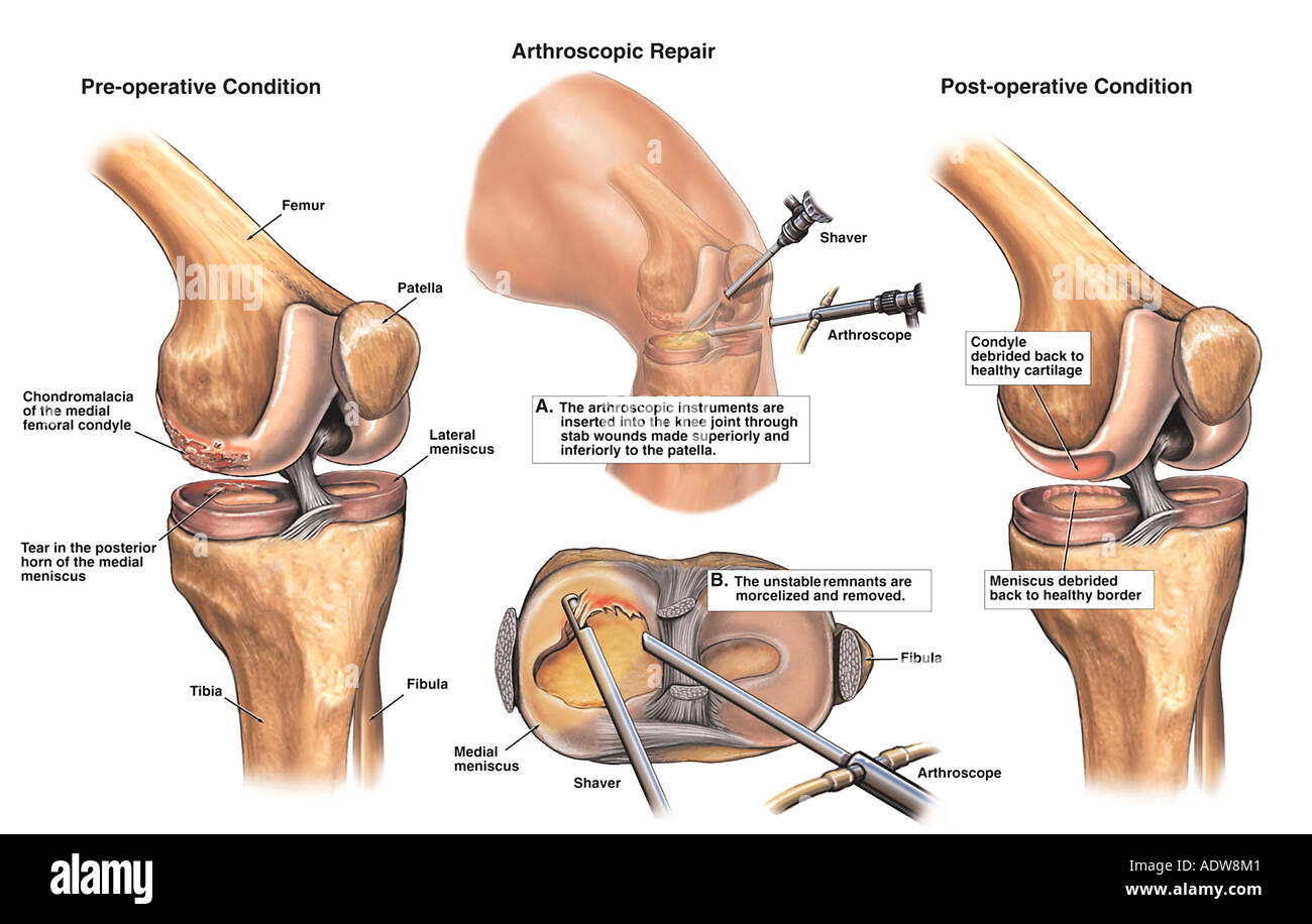
The radiographs demonstrated abnormal contour of the medial femoral condyle, consistent with an osteochondral defect, and a fabella posterior to the knee.
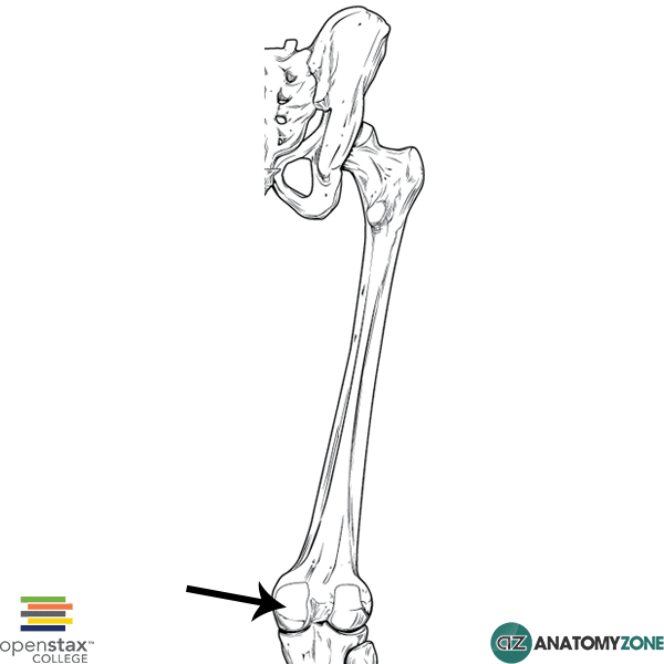
Medial condyle of femur from wikipedia, the free encyclopedia the medial condyle is one of the two projections on the lower extremity of femur, the other being the lateral condyle. The lateral condyle was involved in 44 % and the medial condyle in 12 %. Palpable as a hard, rounded bump to the inside of either knee joint, they are one of two condyles at the bottom of each leg bone, the other being the lateral femoral condyle. This adaptable graft option is most commonly used with oats or shell techniques to restore cartilage in the knee, but can also be used to for cartilage restoration of other joints. In a review of 79 patients with this disease, performed to establish guidelines f … The medial femoral condyle is supplied by a plexus of vessels from the descending genicular artery and the medial superior genicular artery. The medial condyle is larger than the lateral (outer) condyle due to more weight bearing caused by the centre of mass being medial to the knee. Root tear (radial tear) of the medial meniscus mild or moderate knee arthritis According to the hospital for special surgery, the medial femoral condyle is the inside of the knee, and health issues dealing with it can be treated. Osteonecrosis of the medial femoral condyle can be treated in a variety of ways depending on the stage of the disease. The medial condyle is named for its location on the inside of the knee, closer to the midline of the body, while the lateral condyle is found on the outside of the knee, away from the midline of the body. In the knee, chondromalacia is usually related to injury, overuse of the knee, and poorly aligned muscles and bones around the knee joint. Your knee mri will often show a:
Palpable to either side of the knee joint when it is bent, they are known specifically as the medial and lateral femoral condyles. Sagittal plane fracture of the medial femoral condyle. Damage may also be the result of a direct blow to the knee. The flap is marked by identifying the vascular plexus on the medial condyle and incorporating a component of the network in the flap. Medial femoral condyle fracture study results.
Medial Femoral Condyle Anatomyzone from anatomyzone.com Chondral injuries may accompany an injury to a ligament, such as the anterior cruciate ligament. Your knee mri will often show a: Schatzker i tibia plateau fracture. Osteonecrosis of the medial femoral condyle presents as a sudden onset of pain on the medial side of the knee. Normal irregular ossification of the femoral condyles was present in 66% of the boys and 44 % of the girls 1 in a review of knee radiographs of 147 healthy, asymptomatic children between the ages of 3 and 13 years. However, two of the patients had one or more fractures missed when they first presented. A bone fracture at this location is termed a femoral condyle fracture. According to the hospital for special surgery, the medial femoral condyle is the inside of the knee, and health issues dealing with it can be treated.
Cartilage can be focally damaged, producing a pot hole in the joint surface, when the knee ligaments are injured.
Of the six patients who had suffered an isolated fracture of their medial condyle, four of the patients had their fractures diagnosed on the first visit. The lateral condyle was involved in 44 % and the medial condyle in 12 %. It acts to support a significant amount of the patient's body weight. Cartilage can be focally damaged, producing a pot hole in the joint surface, when the knee ligaments are injured. Osteonecrosis of the medial femoral condyle presents as a sudden onset of pain on the medial side of the knee. A bone fracture at this location is termed a femoral condyle fracture. The femoral condyle is a thickened area of the femur just above the knee. One presumed mechanism of injury is a stieda fracture (avulsion injury of the medial collateral ligament at the medial femoral condyle). Root tear (radial tear) of the medial meniscus mild or moderate knee arthritis Coronal plane fracture of the lateral femoral condyle. The lesions were located on the medial femoral condyle in 8 (72.7%) cases and on the medial tibial plateau in 3 cases (27.3%). The medial femoral condyles are the bony protrusions on the inside edge of the bottom of the femur bone in each thigh. Based on the patient's antalgic gait and radiographic findings, the patient was instructed on the proper use of crutches and referred to an orthopaedic surgeon for appropriate management.
The lateral condyle was involved in 44 % and the medial condyle in 12 %. The flap is marked by identifying the vascular plexus on the medial condyle and incorporating a component of the network in the flap. This is associated with a positive bone scan and, frequently, a radiolucent lesion in the subchondral zone. If there is a fracture (break) in part of the condyle, this is known as a fracture of the femoral condyle. Medial condyle of femur from wikipedia, the free encyclopedia the medial condyle is one of the two projections on the lower extremity of femur, the other being the lateral condyle.
Knee Normal Anatomy Dorsal Medial View Stock Illustration 498636940 from image.shutterstock.com Damage may also be the result of a direct blow to the knee. The medial femoral condyles are the bony protrusions on the inside edge of the bottom of the femur bone in each thigh. This is associated with a positive bone scan and, frequently, a radiolucent lesion in the subchondral zone. An imbalance of the muscles around the knee (some muscles are weaker than others.) overuse (repeated bending or twisting) of the knee joint, especially during sports. Methods sixteen knees with a small medial femoral. One presumed mechanism of injury is a stieda fracture (avulsion injury of the medial collateral ligament at the medial femoral condyle). Normal irregular ossification of the femoral condyles was present in 66% of the boys and 44 % of the girls 1 in a review of knee radiographs of 147 healthy, asymptomatic children between the ages of 3 and 13 years. A bone fracture at this location is termed a femoral condyle fracture.
Palpable to either side of the knee joint when it is bent, they are known specifically as the medial and lateral femoral condyles.
Methods sixteen knees with a small medial femoral. The lesions were located on the medial femoral condyle in 8 (72.7%) cases and on the medial tibial plateau in 3 cases (27.3%). Radiographic features it is almost always unilateral, usually affects the medial femoral condyle (but can occasionally involve the tibial plateau 9) and is often associated with a meniscal tear. In the knee, chondromalacia is usually related to injury, overuse of the knee, and poorly aligned muscles and bones around the knee joint. It acts to support a significant amount of the patient's body weight. Normal irregular ossification of the femoral condyles was present in 66% of the boys and 44 % of the girls 1 in a review of knee radiographs of 147 healthy, asymptomatic children between the ages of 3 and 13 years. The inner side of the knee or the medial femoral condyle is the most common area for a cartilage defect. A bone fracture at this location is termed a femoral condyle fracture. Palpable to either side of the knee joint when it is bent, they are known specifically as the medial and lateral femoral condyles. If there is a fracture (break) in part of the condyle, this is known as a fracture of the femoral condyle. Primary osteonecrosis of the femoral condyle shares several features with insufficiency fractures, including predominance in elderly women with factors responsible for mechanical stress (varum, obesity, trivial trauma), mechanical pain, and increased radionuclide uptake. An imbalance of the muscles around the knee (some muscles are weaker than others.) overuse (repeated bending or twisting) of the knee joint, especially during sports. Sagittal plane fracture of the medial femoral condyle.

 in part of the condyle, this is known as a fracture of the femoral condyle.)